Where Is The Trigeminal Ganglion Situated
It is situated above the deep portion of the submandibular gland on the hyoglossus muscle near the posterior border of the mylohyoid muscle. The peripheral aspect of the trigeminal ganglion gives rise to 3 divisions.
 Pin On Migraine Bleaaah
The trigeminal ganglion is located lateral to the cavernous sinus in a depression of the temporal bone.

Where is the trigeminal ganglion situated. A ganglion appears as a swelling along the course of a nerve. The autonomic ganglia of the parasympathetic nervous system. The pterygopalatine ganglion of Meckel the largest of the parasympathetic ganglia associated with the branches of the maxillary nerve is deeply placed in the pterygopalatine fossa close to the sphenopalatine foramenIt is triangular or heart-shaped of a reddish-gray color and is situated just below the maxillary nerve as it crosses the fossa.
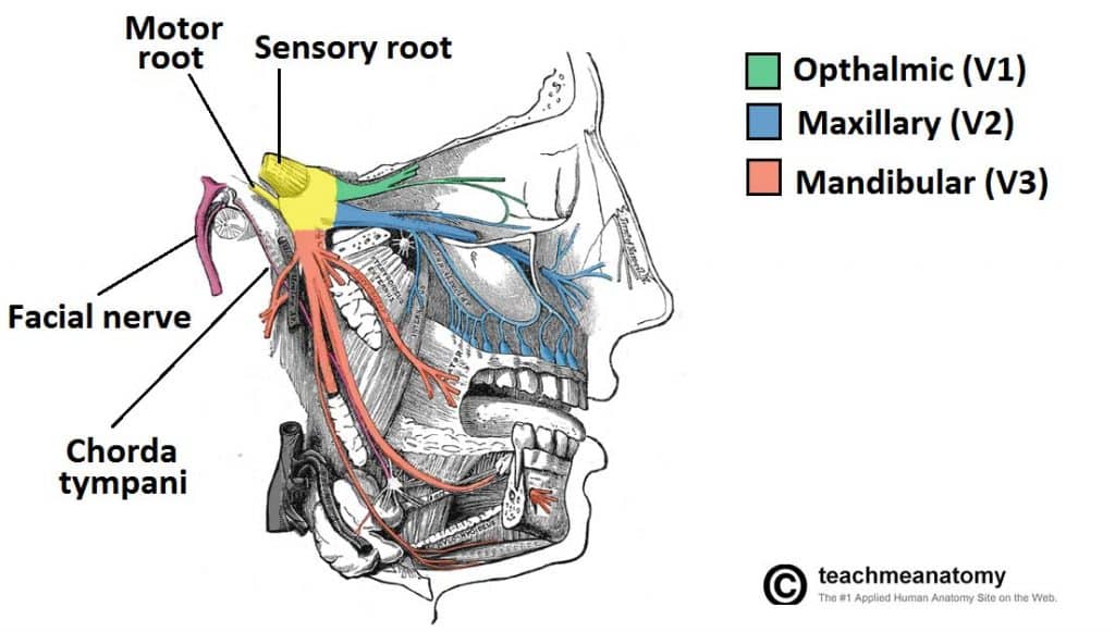
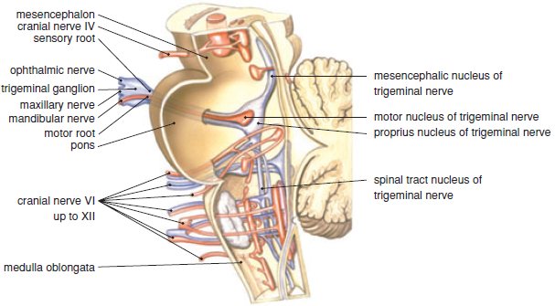
Motor fibres are only distributed to the mandibular division V3. It is situated below the foramen ovale. The Cranial SympatheticsThe cranial sympathetics include sympathetic efferent fibers in the oculomotor facial glossopharyngeal and vagus nerves as well as sympathetic afferent in the last three nerves.
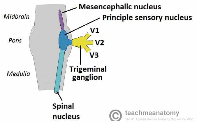
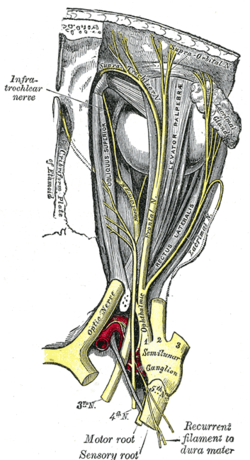
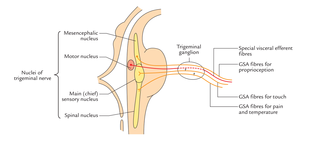
The spinal ganglia or posterior or dorsal root ganglia associated with the spinal nerves contain the unipolar neurons of the sensory nerve fibers that carry signals to the cord. This depression is known as the trigeminal cave. The motor root passes inferiorly to the sensory root along the floor of the trigeminal cave.
The portion of the nerve before the ganglion is referred to as preganglionic and carries the impulse towards the cluster of cell bodies. The precentral gyrus also known as the primary motor cortex is a very important structure involved in executing voluntary motor movements. The ANS has three major branches.
The precentral gyrus is a diagonally oriented cerebral convolution situated in the posterior portion of the frontal lobe. The portion located from the ganglion onwards is called postganglionic and carries the impulse away from the cell bodies. The submandibular ganglion is small and fusiform in shape.
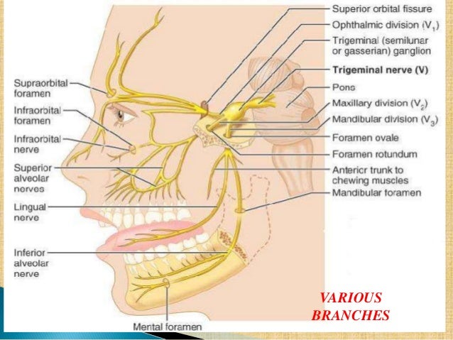
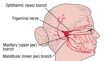
THE TRIGEMINAL GANGLION Sensory root fibres of the trigeminal nervecomprise the centralprocess of cells located in the trigeminalganglion2 ganglion one inervating each side of facelocated inmeckels cavityon the anterior surface of the petrous portion oftemporal bonemeasures arprox. There are 3 divisions of the trigeminal nerve that occur due to its peripheral nature Mandibular Ophthalmic and Maxillary. Ophthalmic V1 maxillary V2 and mandibular V3.
The trigeminal ganglion is located lateral to the cavernous sinus in a depression of the temporal bone known as the trigeminal cave or Meckels cave. The ganglion hangs by two nerve filaments from the lower border of the lingual nerve itself a branch of the mandibular nerve CN V 3. The Sympathetic Efferent Fibers of the Oculomotor Nerve probably arise from cells in the anterior part of the oculomotor nucleus which is located in the tegmentum of the mid-brain.
Its importance lies as the neurovascular crossroad of the nasal cavity masticator space orbit oral cavity and middle. The pterygopalatine fossa PPF less commonly known as the sphenopalatine fossa is a small but complex space of the deep face in the shape of an inverted pyramid located between the maxillary bone anteriorly the pterygoid process posteriorly and orbital apex superiorly. Trigeminal ganglion a ganglion on the sensory root of the fifth cranial nerve situated in a cleft within the dura mater on the anterior surface of the pars petrosa of the temporal bone and giving off the ophthalmic and maxillary and part of the mandibular nerve.
A dorsal root ganglion or spinal ganglion is a nodule on a dorsal root of the spine that contains the cell bodies of nerve cells neurons that carry signals from sensory organs toward the appropriate integration center. It is located immediately anterior to the central sulcus fissure of Rolando running parallel to it 1-2. A ganglion is a cluster of neuron cell bodies enveloped in an epineurium continuous with that of a nerve.
The trigeminal ganglion is situated lateral close to the cavernous sinus forming a depression of the temporal bone which is called as the trigeminal cave. Most are small. Sympathetic parasympathetic and enteric.
 Pin On Anatomy
 Diagram Of The Second Branch Maxillary Of The Tr Nerve Anatomy Cranial Nerves Anatomy Gross Anatomy
Pterygopalatine Fossa Maxillary Nerve Nerve Anatomy Dental Anatomy
 Pin On Neuro
 Olfactory Nerve Function Location Related Conditions And Faqs Nerve Anatomy Nerve Cranial Nerves
 Trigeminal Nerve Anatomy
 The Trigeminal Nerve Cn V Course Divisions Teachmeanatomy
 Peripheral Nervous System Cranial Nerves Optic Nerve Nerve Anatomy Cranial Nerves
 Pin On Neuro
 Abducens Nerve Function Location Anatomy And Faqs Abducens Nerve Cranial Nerves Nerve Anatomy
 The Ophthalmic Division Of The Trigeminal Nerve Cnv1 Teachmeanatomy
 Cambridge Questions Facial Nerve Maxillary Nerve Dental Anatomy
 Pin On Nerves
 Trigeminal Ganglion Wikipedia
 Easy Notes On Trigeminal Nerve Learn In Just 3 Minutes Earth S Lab
 Trigeminal Neuralgia Tic Douloureux Harvard Health
 Peripheral Nervous System Cranial Nerves Vagus Nerve Nerve Anatomy Peripheral Nervous System
 Pin On Eye
 2 Anatomy Of The Trigeminal Nerve Pocket Dentistry