Posteromedial Capsule Knee Mri
In complex knee injuries the patient is positioned supine with both knees in extension. Abstract The medial and posteromedial regions of the knee are important for knee stability but also frequently injured.
 The Total Knee Replacement Attorneys Kassel Mcvey Attorneys At In 2021 Total Knee Replacement Knee Surgery Knee Replacement Surgery
The Posteromedial Corner on MRI part 2 - 5 min.
Posteromedial capsule knee mri. Most common in the posteromedial meniscocapsular interface Ganglia increased meniscocapsular fat and capsulosynovial redundancy can simulate meniscocapsular separation In true separation the meniscus moves anteriorly folds flips extrudes or migrates Separation without associated meniscal tear bone or cartilage or PMC injury is rare6. The lesion was reported to have multiple septae and was closely related to the semimembranosus muscle Figures 3a and 3b. Capsule of the Knee joint Proximal extent follows contour of suprapatellar pouch and articular cartilage Distal extent meniscotibial ligament and articular cartilage Anterior.
MRI allows an accurate assessment of the posterior knee capsule. In the Knee MRI Mastery courses we give you everything you need in order to evaluate this joint. Concise knowledge of these relationships is necessary before approaching their evaluation at imaging.
With modern MRI systems these structures are readily identified and can be appreciated in the context of multiligamentous knee injuries. The findings in 10a-12a indicate a combined PCL injury with associated injury of the posterolateral corner. Posteromedial corner injury of the knee is a readily identifiable but frequently underappreciated injury on imaging.
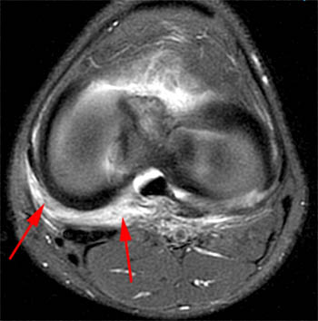
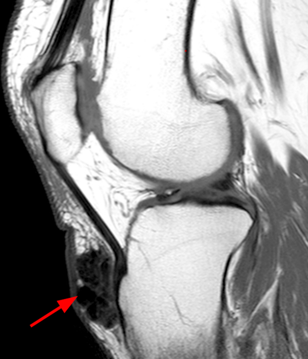
The authors used a three-layer approach to correlate the appearance of the capsule and ligaments of the medial side of the knee on magnetic resonance MR images with corresponding anatomic slices. Hemorrhage and edema are present along the posteromedial capsule and posterior oblique ligaments arrowheads. An MRI of the knee and leg was obtained which revealed a large cystic lesion measuring 24 10 12 cm in the posteromedial aspect of the knee and calf.
Following the MRI an excision was planned. In contradistinction this knee region represents a blind area for the arthroscopist. Intervention to the posteromedial capsule is performed in the appropriate patient based on physical exam preoperative imaging and arthroscopy as an additional procedure to ACLR Table 1.
The Posteromedial Corner on MRI part 3 - 3 min. Posterior Capsule - 4 min. Importantly it can result in increased stress on the cruciate ligaments and can result in anteromedial rotatory instability AMRI of the knee.
Deep fibers of MCL Meniscofemoral ligament Meniscotibial ligament Posterior. MR images of six fresh cadaveric specimens were obtained by using a proton-densityweighted fast spin-echo sequence with a 256 512 matrix. Capsule and patellomeniscal ligament Mid.
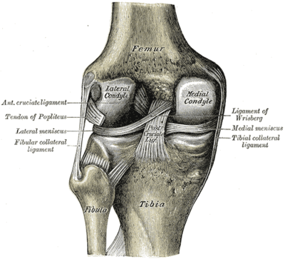
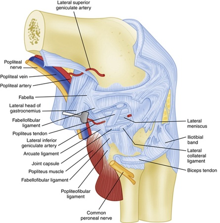
The posteromedial corner of the knee encompasses five medial structures posterior to the medial collateral ligament. MRI readily delineates injuries of bone other ligaments menisci cartilage capsule tendons and vascular structures. However injuries of the posterior capsule are often associated with lesions of the menisci and the cruciate ligaments.
MRI signal of a healing meniscocapsular rupture manifests as low signal meniscocapsular attachment of the PHMM. Medial ligaments and capsule are primary and secondary stabilizers of valgus rotation and anterior and posterior translation. At 3months postoperatively MRI was used to confirm healing of meniscocapsular rupture.
Because MRI is commonly performed in the setting of knee injury radiologists familiar with the normal and abnormal appearances of the posterolateral corner structures on MRI can suggest the diagnosis of posterolateral corner injury when present leading to improvements in treatment and functional outcomes for patients in whom the injury was not clinically suspectedin particular in those patients with concomitant ACL or PCL injuries requiring reconstruction. The Posteromedial Corner on MRI. Posteromedial joint capsule PJC and posterior horn of medial meniscus PHMM1 PJC is composed of meniscotibial and meniscofemoral components and being reinforced by the POL the posteromedial joint capsule extends to the medial head of the gastrocnemius Both PJC and MM Layer III are reinforced externally by POL.
The posteromedial corner of the knee PMC is an important anatomic structure that is easily seen but often overlooked on magnetic resonance MR images. Injury to one of the main structures comprising the posteromedial corner PMC of the knee with modern MRI systems the major anatomic structures comprising the PMC can be readily identified these structures contribute to the static and dynamic stability of the knee including a supporting role in multiligament knee injuries. Posterolateral PLC and posteromedial PMC corners of the knee represent complex anatomic regions because of intricate soft tissue and osseous relationships in small areas.
The posterior oblique ligament POL belongs to the medial supporting structures of the knee and is one of the five components of the posteromedial corner PMC of the knee. The posterior capsule may be injured in hyperextension trauma. Improved knee function score and no posteromedial knee pain or flexionsquatting limitation were also confirmed.
The posteromedial corner of the knee PMC is comprised of the structures between the posterior border of the superficial medial collateral ligament SMCL and the medial border of the posterior cruciate ligament PCL. It is recognized that anteromedial rotatory i. We can be your guide through the intricacies of the knee.
 Posterolateral Corner Injury Of The Knee Radiology Reference Article Radiopaedia Org
 Posteromedial Corner Injury Of The Knee Radsource
 Pin On Health
 Oblique Popliteal Ligament Radiology Reference Article Radiopaedia Org
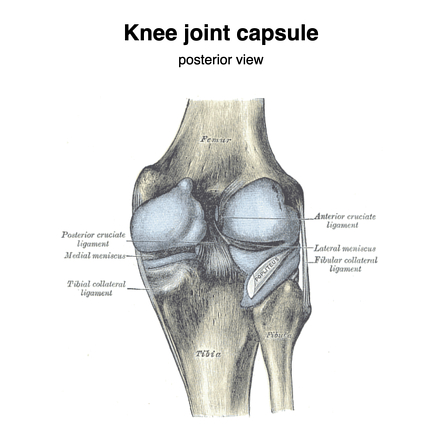
 Posterior View Of The Knee With Posterior Capsules Shown In Blue Download Scientific Diagram
 Pin On Bones Muscles
 Anatomy Knee Knee Joint Anatomy Anatomy Of The Knee Bursitis Knee
 Knee Capsule Radiology Reference Article Radiopaedia Org
 Pin On Anatomy
 Posterolateral And Posteromedial Corner Injuries Of The Knee Radiology Key
Https Www Clinicalradiologyonline Net Article S0009 9260 07 00061 X Pdf
 Muscles Anatomy Physiology Health Fitness Training Muscle Bone Knee Medical Anatomy Muscle Anatomy Body Anatomy
 Ligaments During Knee Flexion And Extension Human Anatomy And Physiology Kinesiology Biomechanics
 Knee Bursae Radsource
 Viewing Playlist Aa Junepin Radiopaedia Org Radiology Nuclear Medicine Osteopathy
 Posterolateral Corner Plc Knee Injuries
 Complex Meniscus Tear Bing Images Meniscus Tear Knee Knee Injury
 Pin By Chyenne Thorpe On Awesome Stuff In 2021 Baker S Cyst Swollen Knee Cysts
Medial Collateral Ligament Of The Knee Physiopedia